The neural circuit basis for motor learning tasks when myelination is impaired has been illuminated for the first time by an international collaboration of university research teams. They also succeeded in compensating for the impaired motor learning process by pairing appropriate actions with brain photo-simulation to promote synchronization of neuronal activities. This could contribute to future treatments for neurological and psychiatric diseases in which white matter function is impaired.
The research was carried out by Assistant Professor Daisuke Kato and Professor Hiroaki Wake (Kobe University Graduate School of Medicine, Japan), Professor Junichi Nabekura (National Institute of Physiological Sciences, Japan), Dr. R Douglas Fields (National Institutes of Health, USA) and Professor Masanori Matsuzaki (Tokyo University Graduate School of Medicine, Japan).
The results were first published in the journal ‘GLIA’.
Introduction
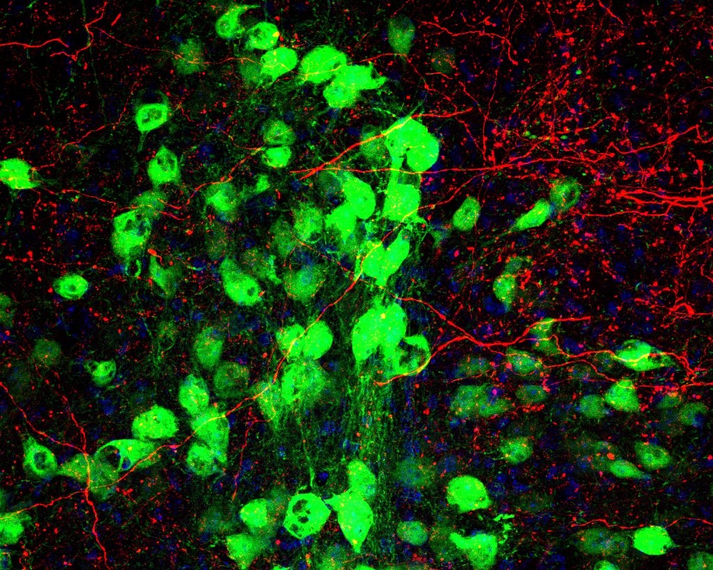
Myelin is sheath that forms around axons, regulating the speed of electrical impulses and efficiently transmitting them among the neurons. Myelinated bundles act as cables to connect distant brain regions. Once myelination is impaired or the myelin is damaged, the propagation of impulses in the neurons slows down or is dysregulated. This impaired regulation has been linked to abnormal activity in neuronal populations, resulting in learning deficits and aging (particularly in dementia and Alzheimer’s disease). The resulting changes in white matter have been observed in the MRI scans of patients with Alzheimer’s. However, it is still poorly understood how exactly impaired myelination affects the circuit properties of the brain that are important for learning and cognition.
This research showed that impaired myelination causes uncoordinated or asynchronous electrical impulse transmission between neurons. Impaired myelination was shown to have an adverse effect on motor learning in mice, suggesting that coordinated transmissions are vital for effective learning.
Research methodology
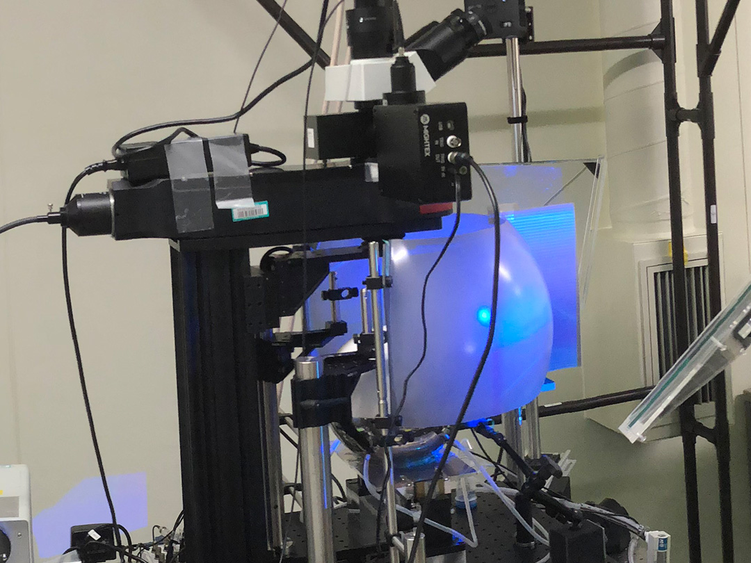
The population activity of neurons in the primary motor cortex of mice with myelin impairments was measured during a motor learning activity using in vivo two photon microscopy. Mice with head plates were inserted into body chambers. The mice were trained to pull and hold a lever that would dispense drops of water. Mouse behavior was monitored by infrared video camera. In the early stages of training there was no difference in the performance between myelin impaired mice and control mice. However, in the later stage of training the myelin impaired mice had a lower success rate in performing the task, although the amount of attempts was similar. Although this suggests their motivation levels were the same, the myelin deficit made it more difficult for the mice improve their performance of this task. It also reduced the accuracy of their movements and increased the spontaneous activities of neuronal population.
Through analyzing the activity of the neurons of myelin impaired mice, they showed that asynchronous activity in the thalamocortical axons correlated with impaired task performance. Thalamocortical axons are nerve fibers connecting the thalamus and cerebral cortex of the brain which carry nerve cells’ information. Electrical stimulation of the motor cortex (output area) during the lever pull task was utilized to promote synchronous activity of neurons in motor cortex and to try to compensate for the performance of the mice. This promoted synchronous activity in the thalamocortical axons during learning and improved the success rate of the mice with myelin impairment.
Conclusions
The findings of this research illuminate how pathological neuronal circuit activity is affected by impaired myelination. The results also suggest that it may be possible to pair noninvasive brain simulation with relevant behaviors to correct cognitive and behavioral abnormalities in the early stages of diseases with impaired white matter.
Glossary
- Thalamocortical axons
- are nerve fibers connecting the thalamus and cerebral cortex of the brain which carry nerve cells’ information.
- Oligodendrocytes
- are specialized brain cells responsible for the myelination in the central nervous system. They are vital to neuron survival.
Acknowledgements
This research was supported by MEXT (Ministry of Education, Culture, Sports, Science and Technology), JST (Japan Science and Technology Agency) and AMED (Japan Agency for Medical Research and Development).
Journal information
- Title
- “Motor learning requires myelination to reduce asynchrony and spontaneity in neural activity”
- DOI
- 10.1002/glia.23713
- Authors
- Daisuke Kato, Hiroaki Wake, Philip R Lee, Yoshihisa Tachibana, Riho Ono, Shouta Sugio, Yukio Tsuji, Yasuyo H Tanaka, Yasuhiro R Tanaka, Yoshito Masamizu, Riichiro Hira, Andrew J Moorhouse, Nobuaki Tamamaki, Kazuhiro Ikenaka, Noriyuki Matsukawa, R Douglas Fields, Junichi Nabekura, and Masanori Matsuzaki
- Journal
- GLIA