Project Description
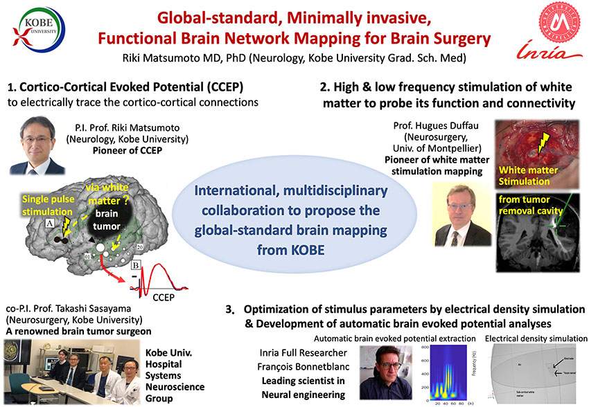
The brain is an important organ that orchestrates various higher functions that make us human, such as motor and sensory functions, and those related to language, memory, emotion and cognition. Brain surgery is a treatment option for brain diseases such as brain tumors and epilepsy. It is crucial to preserve these important functions during surgery so that the patient will have better quality of life afterwards. Direct electrical cortical stimulation has been the standard method to map cortical functions for brain surgery since the mid-20th century, but has potential adverse effects such as the risk of inducing convulsions. Furthermore, it is not possible to map the whole network of a particular area of the brain, namely, the functionally relevant cortical regions and the white matter pathways that connect these regions.
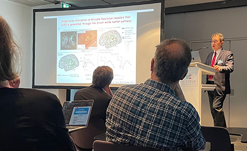
Professor MATSUMOTO Riki (Professor at the Graduate School of Medicine) has developed a unique method called cortico-cortical evoked potential (CCEP) to electrically trace the cortico-cortical networks by injecting minimal electric currents into the cortex. Based on Prof. MATSUMOTO’s cutting-edge research outcomes, the international joint research project ‘Development of a global-standard, minimally invasive, functional brain network mapping technique for brain surgery’ was started in 2022. This three-year project has been awarded the Kobe University Strategic International Collaborative Research Grant (Type B: Fostering Joint Research). In this project, we promote international multidisciplinary collaboration, primarily with two French research institutions; 1 ) the University of Montpellier team led by Professor Hugues Duffau, who pioneered the electrical stimulation of white matter to map the function of white matter fibers, and 2) the Inria team led by Dr. François Bonnetblanc, a renowned expert in analyzing brain evoked potentials and electrostimulation. The Kobe University Hospital Systems Neuroscience Group (led by Professors MATSUMOTO (Neurology) and SASAYAMA Takashi (Neurosurgery)), incorporate the state-of-art expertise and techniques of the French-Japanese team to research and develop a minimally invasive, functional brain ‘network’ mapping technique for brain surgery that can become the new global-standard.
Comments from the Project Leader
Professor MATSUMOTO Riki (Professor of the Graduate School of Medicine)
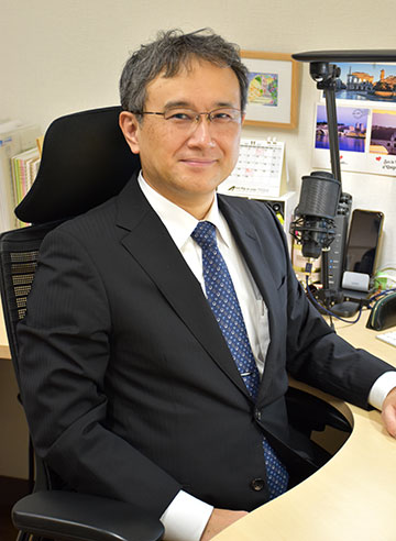
Our brain can be compared to a small universe. Throughout my career as a clinical neurophysiologist responsible for clinical functional brain mapping, I have felt that untangling its mysteries requires world-class expertise to improve the state-of-the-art systems neuroscience we provide to patients undergoing neurosurgery. In this project, we would like to promote French-Japanese collaboration for the development of a global-standard, minimally invasive, functional brain network mapping technique as the project title illustrates. We believe the establishment of our mapping method will lead to better functional outcomes for patients after brain surgery.
Our brain can be compared to a small universe. Throughout my career as a clinical neurophysiologist responsible for clinical functional brain mapping, I have felt that untangling its mysteries requires world-class expertise to improve the state-of-the-art systems neuroscience we provide to patients undergoing neurosurgery. In this project, we would like to promote French-Japanese collaboration for the development of a global-standard, minimally invasive, functional brain network mapping technique as the project title illustrates. We believe the establishment of our mapping method will lead to better functional outcomes for patients after brain surgery.
International Collaboration
Through the project, we have launched various collaborative activities that we plan to conduct over the next three years.
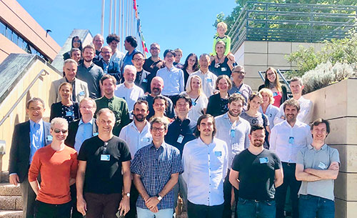
In June 2022, we held an international kick-off workshop entitled “Workshop on Brain Evoked Potentials in the context of Neurosurgery” in Montpellier, France. The Kobe group had the opportunity to visit Prof. Duffau at the University Hospital to learn about state-of-art white matter mapping during awake surgery. We also had an on-site collaborative meeting at Montpellier branch of Inria. Since then, we have been having regular online meetings. The Montpellier team plans to visit us in March 2023 to refine our new mapping method. In the fiscal year 2023, the Kobe team plans to visit Montpellier and Paris for further collaboration.

Information on related research facilities
Riki Matsumoto (P.I.)
Takashi Sasayama (co-P.I.)