The membranes of cancer cells are more pliant than the membranes of normal cells. A research collaboration has discovered that cancer invasion and migration can be supressed in mice by manipulating the stiffness of the cell membrane. The research group included Lecturer TSUJITA Kazuya and Professor ITOH Toshiki, both of Kobe University’s Biosignal Research Center, and Lecturer SATOW Reiko and Professor Emeritus FUKAMI Kiyoko from Tokyo University of Pharmacy and Life Sciences.
It is hoped that this finding can be applied to the development of novel cancer treatments that exploit the physical characteristics of cells.
These research results were published in Nature Communications on October 11, 2021.
Main points
- Cancer cell membranes are softer compared to those of normal cells.
- By increasing the membrane tension of cancer cells, the research group succeeded in supressing their migration and invasion in a mouse model.
- This will contribute towards new treatments that target the physical characteristics of cancer cells.
Research Background
Metastasis is the major cause of cancer-related deaths. As a cancer cell’s malignancy increases, it undergoes amoeba-like structural changes that enable it to migrate more easily. It moves away from the primary lesion, triggering distant metastasis. In recent years, research has revealed that these significant changes to cellular structure and motility are controlled by the physical characteristics of the cell itself. In fact, it has been reported that cancer cells are comparatively ‘soft’ compared to normal cells. The connection between changes in a cell’s physical characteristics and its malignancy has received much attention. However, is not known exactly which physical characteristics are related to a cell’s cancerous nature.
Research Findings
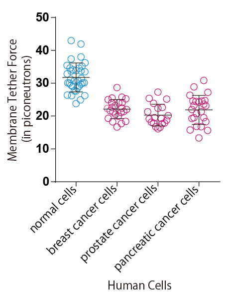
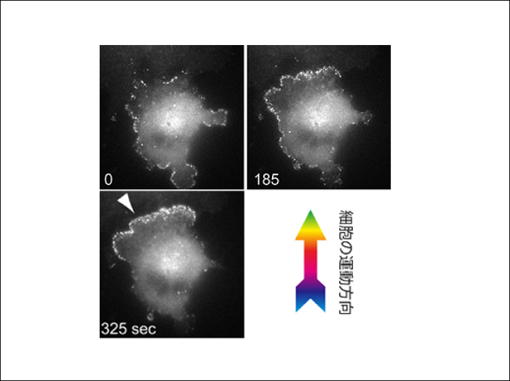
In their research, Tsujita et al. used optical tweezers* to pull the cell surface membrane and analyse it, which enabled them to determine that cancer cells are comparatively softer than normal cells (Figure 1). The firmness of the cell membrane is regulated by the actin cytoskeletal networks that attach to it. This study revealed that in cancer cells, the ERM proteins that maintain this membrane-actin attachment are dissociated from the cell membrane, which makes the membrane soft.
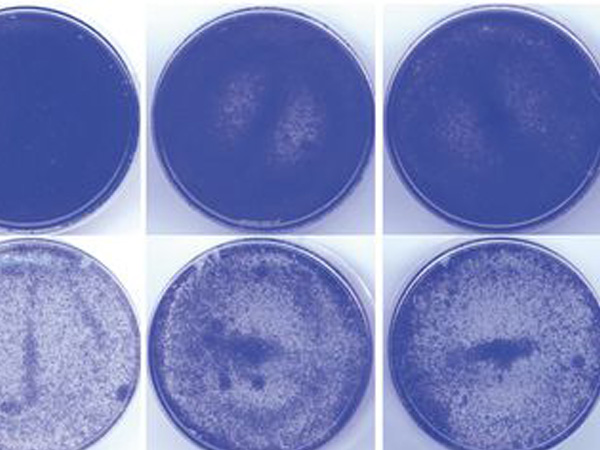
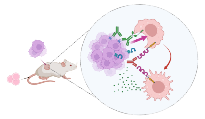
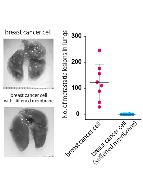
By securely attaching ERM proteins to cancer cell membranes, the researchers were able to restore the membrane-actin attachment so it resembled that of a normal cell. This caused the cancer cell membrane to stiffen and prevented abnormal changes in structure and motility. Furthermore, breast cancer cells with stiffened membranes lost the ability to spread to the lungs in experiments using a mouse model (Figure 2). These results indicate that it could be possible constrain the spread of cancer by manipulating cell membrane tension.
Further Developments
The findings of the current research study could lead to the development of new cancer treatments that exploit the physical characteristics of cancer cells. If the chemical compound that stiffens cell membranes is discovered, then it may also be possible to utilize this in effective medication to prevent cancer invasion and migration.
Glossary
- *Optical tweezers
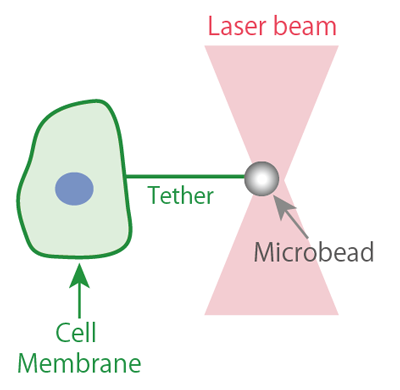
- Optical tweezers are concentrated beams of laser light that can be used to trap and move microbeads. Attaching these trapped microbeads to the cell membrane and pulling them forms a membrane tether (a string-like structure) enabling the cell membrane tension to be measured. This functions like a microscopic spring gauge. In 2018, the development of the optical tweezers was awarded the Nobel Prize in Physics.
Acknowledgements
This research was supported by an AMED-PRIME grant from the Japan Agency for Medical Research and Development (Title: ‘Elucidating the mechanism of plasma membrane tension medicated signal transduction in cancer cell invasion and metastasis’ Principle researcher: Tsujita Kazuya). It also received funding from the following Japan Society for the Promotion of Science grants: Grant-in-Aid for Challenging Exploratory Research, Grant-in-Aid for Young Scientists (A), Grant-in-Aid for Scientific Research (B), and a Grant-in-Aid for Scientific Research on Innovative Areas.
Journal Information
- Title
- “Homeostatic membrane tension constrains cancer cell dissemination by counteracting BAR protein assembly”
- DOI
- 10.1038/s41467-021-26156-4
- Authors
- Kazuya Tsujita1,2,3, Reiko Satow4, Shinobu Asada4, Yoshikazu Nakamura4,5, Luis Arnes6, Keisuke Sako7, Yasuyuki Fujita8, Kiyoko Fukami4, and Toshiki Itoh1,2
- Biosignal Research Center, Kobe University, Kobe, Hyogo, 657-8501, Japan.
- Department of Biochemistry and Molecular Biology, Kobe University Graduate School of Medicine
- AMED-PRIME, Japan Agency for Medical Research and Development
- Laboratory of Genome and Biosignals, Tokyo University of Pharmacy and Life Sciences
- Department of Applied Biological Science, Faculty of Science and Technology, Tokyo University of Science
- The Novo Nordisk Foundation Center for Stem Cell Biology (DanStem), Biotech Research & Innovation Centre, University of Copenhagen
- National Cerebral and Cardiovascular Center Research Institute
- Division of Molecular Oncology, Graduate School of Medicine, Kyoto University, Kyoto 606-8501, Japan.
- Journal
- Nature Communications