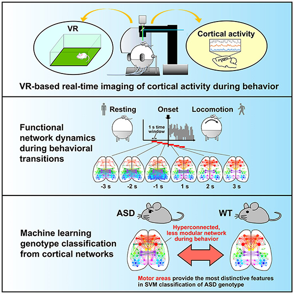
An international research collaboration has developed a VR*1 imaging system that can measure a wide range of neural activity in the cortices of mice during active behavior. This enabled them to illuminate the abnormalities in cortical functional network*2 dynamics that are found in autism*3 model mice. Using machine learning*4, they were also able to highly accurately distinguish between autism model mice and wild-type mice based on the cortical functional network patterns when the mice start or stop running. The research group was led by Professor Toru Takumi and Assistant Professor Nobuhiro Nakai (both of the Department of Physiology, Kobe University Graduate School of Medicine), and Masaaki Sato, (Lecturer, Department of Pharmacology, Graduate School of Medicine, Hokkaido University). Professor Takumi is also a Visiting Senior Scientist at RIKEN Center for Biosystems Dynamics Research.
Future research on functional brain network dynamics in autism is expected to lead to the development of new biomarkers for autism diagnosis.
The results of this research was published in Cell Reports on March 28 at 11am (ET).
Main points
- The researchers developed a VR imaging system that can measure a wide range of cortical activity from mice in action.
- Mice models of autism have dense cortical functional networks and reduced modularity*5 after motor initiation.
- Machine learning can highly accurately identify autism model mice from their cortical functional network patterns.
Research Background
Autism (autism spectrum disorder) is a neurodevelopmental disorder with many unexplored aspects, characterized by poor social communication, intense preoccupation with certain things, and repetitive behaviors. The number of autistic individuals is markedly increasing, which is considered to be a significant social issue. Even now, autism diagnosis is based on behavioral characteristics, which is far from a quantitative perspective, and there is great demand for the discovery of a new biomarker.
In recent years, research has been conducted to identify functional brain abnormalities unique to autistic individuals. Resting-state fMRI*6 studies suggest that the density of functional brain networks increases in young autistic individuals and decreases in adults (Ref. 1). However, these changes vary widely from individual to individual. As the analysis was conducted when the participants were in a resting state, it was unclear how abnormalities in functional brain networks affect behavior.
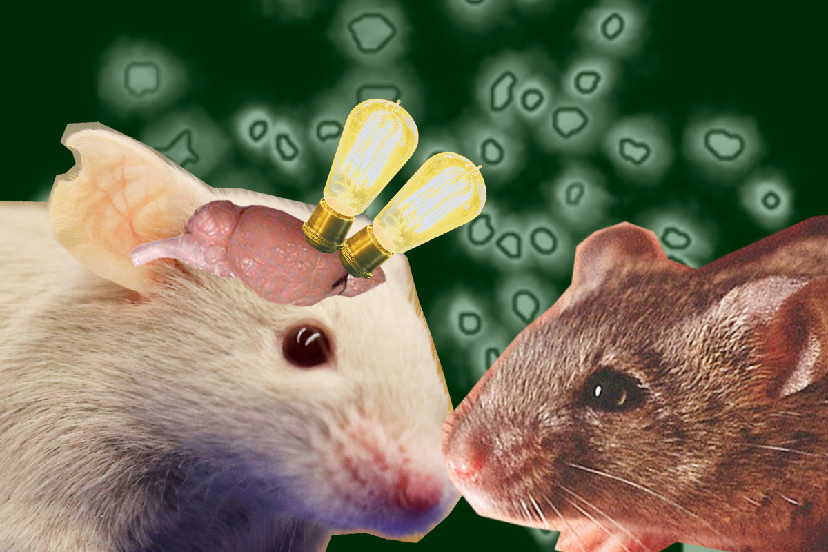
Genetics contribute significantly to autism, and genomic abnormalities such as copy number variations (CNV)*7 are thought to be involved in neuropathology. Recently, animals (mainly mice) modeling human genomic aberrations are often used to elucidate the neuropathology of autism. In this study, the researchers developed a VR imaging system that can measure the brain activity of autism model mice in real-time during active behavior. By investigating brain functional network dynamics, the research group aimed to clarify autism-specific phenomena in the brain during behavior.
Results
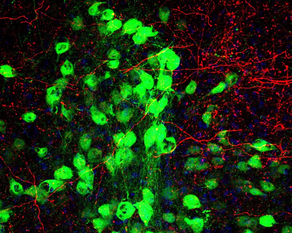
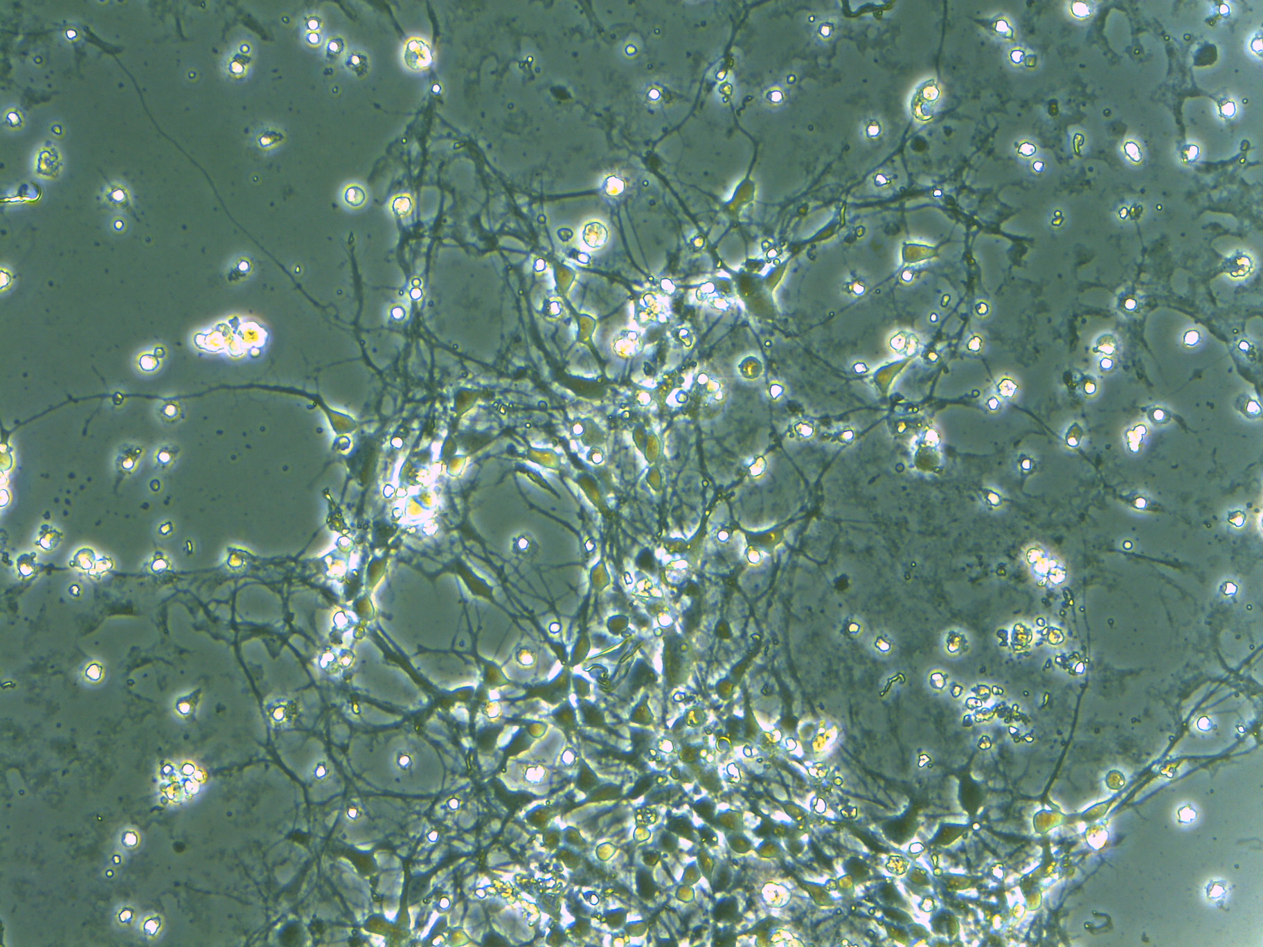
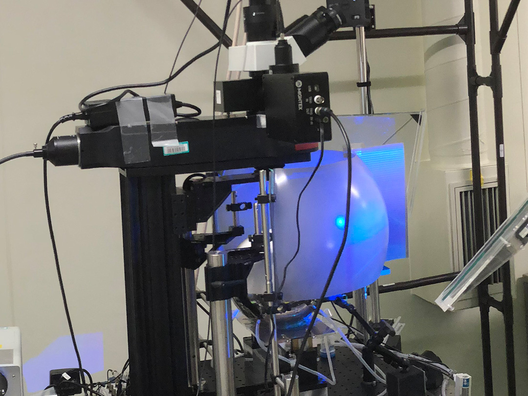
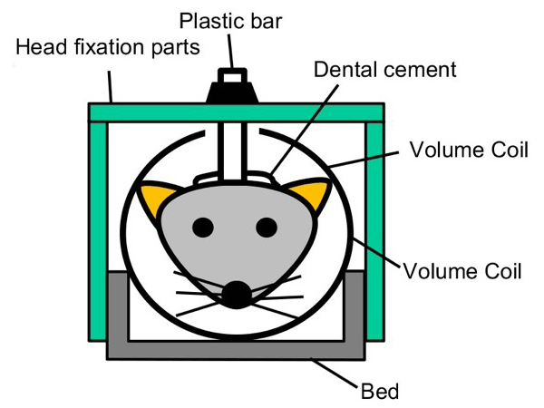
First, a VR imaging system was constructed (Fig. 1A). A mouse with its head fixed in place is put on a treadmill and shown an image of a virtual space projected on a screen. The virtual space was prepared so that it reproduced the field used for mouse behavioral experiments. The motion of the treadmill is reflected in the video images, allowing the mice to freely explore the virtual space (Fig. 1B). Alongside behavioral measurements such as locomotion, transcranial calcium imaging*8 was performed simultaneously so that a wide range of functional area activity in the cerebral cortex could be measured in real time (Fig. 1C-E). For this purpose, the researchers used transgenic mice that express calcium sensor protein (GCaMP)*9 in their neurons. In addition, they established a method for analyzing cortical functional network dynamics. They calculated correlations between functional areas from one-second neural activity data obtained via calcium imaging, and visualized the functional network using graph theory (Fig. 1E).
The researchers analyzed the 3 second time windows before and after when the mouse spontaneously started or stopped moving on the treadmill (locomotion) and examined the network characteristics in each time window. The results revealed that the network structure changes with the onset of locomotion and that modularity increases (Fig. 2). It was also found that the network structure returns to the resting state when locomotion is stopped. Thus, they succeeded in visualizing the network dynamics during the switch from rest to locomotion and from locomotion to rest.
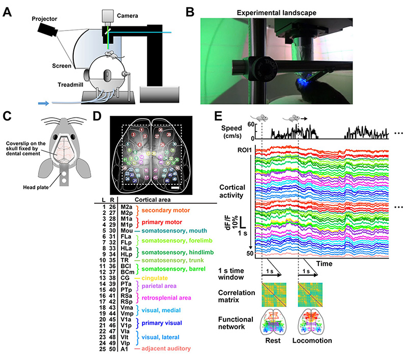
A, The imaging and virtual reality (VR) system.
B, Photograph of the experiment
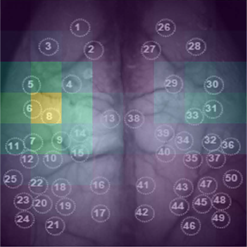
C, A schematic of the transcranial imaging window affixed to the mouse skull.
D, Fifty cortical ROIs are overlaid onto a grayscale image of the dorsal cortex with a cortical parcellation map (top, dashed lines indicate the field of view, scale bar = 1 mm). ROIs 1–25 and 26–50 were defined in the left (L) and right (R) hemispheres, respectively, and ROIs for each hemisphere were numbered along the anterior-posterior axis (bottom).
E, Analysis of cortical functional connectivity.
After calculating normalized fluorescence changes (dF/F) for each ROI, pair-wise Pearson’s correction coefficients of cortical activity in a one-second time window were calculated for all ROI pairs and then visualized as matrices. Each matrix was labeled with a corresponding behavior state at the first frame of the time window. In the graph visualization of functional networks, connectivity with a correlation coefficient above a threshold (r > 0.8) was denoted as a line (edge) that connected the corresponding ROIs (nodes).
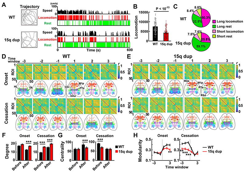
A, Representative trajectory (left) and locomotion behavior (right) for Emx1G6 mice and Emx1G615q dup mice. Locomotion speed and periods of locomotion (Loco) and rest of each genotype are shown from top to bottom in the right panel.
B, Locomotor activity of Emx1G6 mice and Emx1G615q dup mice during 10-min sessions. Data represent mean ± SEM. P-value by t-test. n = 89 sessions from 7 Emx1G6 mice and 88 sessions from 9 Emx1G615q dup mice.
C, Percentages of time spent for each type of episode in Emx1G6 mice and Emx1G615q dup mice. Data represent averages across all sessions.
D-E, Changes in cortical functional network dynamics near the locomotion start and stop points in wild-type (D) and 15q dup mice (E).
F, The average number of functional connections that each region of interest has before and after the behavior change point.
G, Mean value of network centrality for each region of interest.
H, Comparison of Modularity
Next, the researchers used this VR imaging system to analyze the functional cortical network of autism model mice. For the experiment, they used 15q dup mice*10, the first established mouse model of autism with copy number variations. 15q dup mice exhibited reduced locomotion and distance traveled in VR space (Fig. 2A-C). Examination of the functional cortical network revealed higher network connections after locomotion initiation, decreased network centrality, and decreased modularity of the functional network (Fig. 2D-I).
Based on these differences in network patterns, the researchers attempted to identify autism model mice by cortical function networks using support vector machines (SVM), a type of machine learning (Fig. 3A). The network patterns of multiple individual 15q dup mice and wild-type mice were used as a training data and the SVM was able to distinguish whether individual test data was from an autism model mouse or not with 78~89% accuracy rate (Fig. 3B). This result suggests that the functional brain network during behavior contains versatile information about the genotype identification. The researchers also examined which information was influential in the brain and found that functional connectivity in the motor cortex was essential for identification in autism model mice (Fig. 3C).
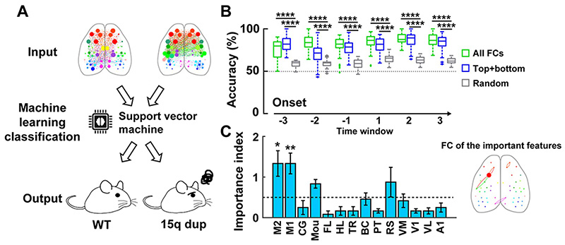
A, The support vector machine learns cortical functional network information before and after the onset of behavior, and discriminates between autism model mice and wild-type mice based on test data.
B, Discrimination accuracy of machine learning when using data from each 1-second time window at the locomotion start point (green: results when trained with all functional bonds, blue: results when trained with only the top 1% functional bonds in the importance index, gray: results when trained with random data).
C, Importance index of functional domains. The higher the value, the more important it is for discriminating between autism model mice and wild-type mice. The figure on the right is a visualization of the most important index connections.
In summary, the 15q dup mice, a model of autism, had a dense functional cortical network during locomotion and reduced modularity. The researchers also found that machine learning can identify autism model mice in a highly accurate manner based on their functional cortical network patterns that are associated with behavioral changes.
Further Research
The functional brain network in mouse models of autism is characterized by the functional connectivity of the motor cortex, which is crucial for determining autism. Detailed studies of these anatomical connections and neurophysiology will help elucidate which networks between the motor cortex and other brain regions play critical roles in autism pathology. In addition, further research on the functional brain network dynamics of autism during active behavior is expected to lead to the discovery of new biomarkers for the diagnosis of autism.
By analyzing the extensive cortical activity recorded from active mice, the researchers were able to visualize the dynamic behavior-dependent changes in the functional cortical network of the brain. VR allows for the creation of multimodal environments that utilize multiple sensory information, including visual, auditory, and olfactory senses. Since a significant symptom of autism in people is impaired social communication, the researchers would like to construct a social environment for mice in the virtual space and investigate how the functional network dynamics change when autism model mice perform social behaviors.
Glossary
- *1 VR (Virtual Reality)
- A 3D space created by a computer. In VR for humans, images are viewed through a headset, etc. In VR for mice, a virtual space is projected onto a dome-shaped screen to provide an immersive experience. Mice can freely explore the virtual space by walking on a treadmill.
- *2 Autism (Autism Spectrum Disorders)
- Autism is a neurodevelopmental disorder whose main behavioral characteristics include impaired social communication and interaction, and repetitive behaviors. Although various types of genetic mutations and genomic abnormalities have been reported in autistic individuals, the cause in many autism cases remains unknown.
- *3 Functional Network
- A network represented by a functional linkage derived from time-series changes in activity between two regions. When two regions show strongly synchronized (highly correlated) activity with each other, the functional coupling is strong. Graph theory is one of the methods to visualize functional brain networks. The network structure can be shown by graphing nodes (brain regions) and edges (functional connections) that cross the correlation threshold.
- *4 Machine Learning
- A technology in which a machine (computer) learns from a large amount of data (training data) to find rules and patterns in the background of the data to predict and classify unknown data (test data). It is classified into supervised learning, unsupervised learning, and reinforcement learning. The support vector machine used in this study is trained through supervised learning, which is used for many classification problems such as speech and image recognition.
- *5 Modularity
- Modules are groups (clusters) that form part of a network. They are categorized by similarity and connectivity. Modularity refers to how many modules a node (brain region) in a network can be divided into.
- *6 fMRI (functional magnetic resonance imaging)
- A method to measure regional cerebral blood flow changes associated with neural activity. Although the temporal resolution is low, it can comprehensively measure the activity of the entire brain.
- *7 Copy number variation
- A deletion or duplication of genomic DNA spanning more than 1 kb on a chromosome. Can be a genetic cause of a disease if it contains a gene implicated in the disease.
- *8 Transcranial Calcium Imaging
- Calcium imaging is a technique to optically measure neural activity-dependent calcium concentration changes in cells and tissues. It has a higher temporal resolution than fMRI because measurements can be made in tens of milliseconds. It is also less invasive for the brain because it measures neural activity through the skull (transcranial), eliminating the need to remove the skull.
- *9 Calcium sensor protein (GCaMP)
- A protein used to detect intracellular calcium ion concentrations via fluorescence signals, GCaMP is a genetically engineered protein probe that combines green fluorescent protein, calmodulin, and a myosin light chain fragment. The binding of calcium ions increases the fluorescence intensity of GCaMP. Since the intracellular calcium concentration increases when neurons are active, neural activity can be also detected as a change in GCaMP fluorescence intensity.
- *10 15q dup mice
- A mouse model that recapitulates the human chromosome 15q11-13 duplication found at high frequency in patients with autism. 15q dup mice exhibit impaired social behavior. Previous studies have identified abnormalities in cortical synaptic connections and the serotonergic nervous system, as well as reduced functional connectivity throughout the brain at rest, but have not examined how brain function is altered when mice exhibit active behavior. In this study, the researchers cross-bred GCaMP transgenic mice and 15q dup mice so that they could use a VR imaging system to analyze brain activity of autism model mice during active behavior.
Acknowledgements
This research was supported by a Grant-in-Aid for Scientific Research (S) from the Japan Society for the Promotion of Science (JSPS), the Japan Science and Technology Agency Moon Shot Type Research and Development Project, Goal 9, and the Takeda Science Foundation Research Grant.
Reference:
- Uddin LQ, Supekar K, Menon V. Reconceptualizing functional brain connectivity in autism from a developmental perspective Front Hum Neurosci. 2013, 7, 458.
Journal Information
- Title
- “Virtual reality-based real-time imaging reveals abnormal cortical dynamics during behavioral transitions in a mouse model of autism”
- DOI
- 10.1016/j.celrep.2023.112258
- Authors
- Nobuhiro Nakai1,2, Masaaki Sato1,3*, Okito Yamashita4,5, Yukiko Sekine1, Xiaochen Fu1, Junichi Nakai6, Andrew Zalesky7, Toru Takumi1,2,8,9*
1 RIKEN Brain Science Institute, Wako
2 Department of Physiology and Cell Biology, Kobe University School of Medicine
3 Department of Neuropharmacology, Hokkaido University Graduate School of Medicine
4 RIKEN Center for Advanced Intelligence Project
5 Department of Computational Brain Imaging, ATR Neural Information Analysis Laboratories
6 Division of Oral Physiology, Department of Disease Management Dentistry, Tohoku University Graduate School of Dentistry
7 Melbourne Neuropsychiatry Centre and Department of Biomedical Engineering, The University of Melbourne
8 RIKEN Center for Biosystems Dynamics Research
9 Lead contact
*Corresponding authors - Journal
- Cell Reports