A research group led by Professor NISHIMURA Noriyuki (Graduate School of Health Sciences, Kobe University) has developed a new method to monitor the residual disease after treatment in high-risk neuroblastoma patients. The method could be utilized to evaluate treatment response and facilitate early diagnosis of tumor relapse/regrowth.
This monitoring method was developed by a group consisting of Assistant Professor YAMAMOTO Nobuyuki and Project Assistant Professor UEMURA Suguru (both of the Graduate School of Medicine, Kobe University), and from Hyogo Prefectural Kobe Children’s Hospital; Vice Director Dr. KOSAKA Yoshiyuki, Department Head Dr. HASEGAWA Daiichiro and chief physician Dr. ISHIDA Toshiaki. Research collaboration and support was also provided by SYSMEX.
These results were first published in the ‘Journal of Molecular Diagnostics’ on December 11, 2019.
Main Points
- Disappearance/inactivation of residual disease after treatment (minimal residual disease: MRD) is essential for sustaining treatment effectiveness and ensuring the long-term survival of high-risk neuroblastoma patients. However, it has been difficult to monitor these changes in MRD using previous methods.
- This research group developed a method for monitoring MRD by detecting the levels of 7 neuroblastoma-associated markers using droplet digital PCR (polymerase chain reaction) (*1).
- This monitoring method enabled a precise monitoring of MRD and an early diagnosis of tumor relapse/regrowth in high-risk neuroblastoma patients.
- This new method will enable the stratification of high-risk neuroblastoma patients and is expected to lead to the development of new treatment methods and improved patient prognoses.
Research Background
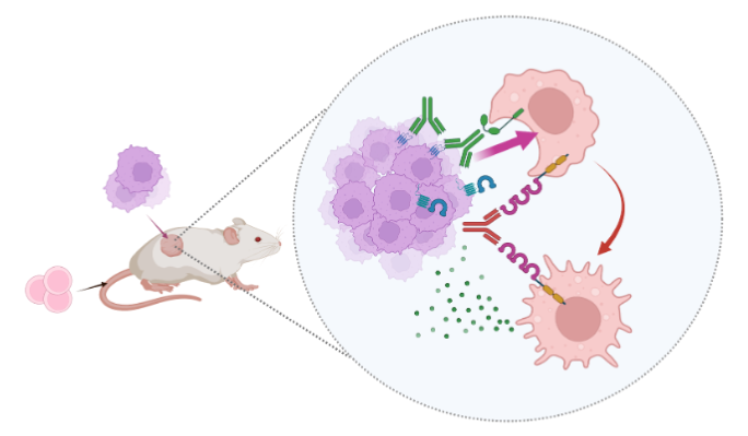
Neuroblastoma is a type of cancer that affects children. It is characterized by solid tumors that develop from immature nerve cells in the sympathetic nervous system, commonly in and around the adrenal glands. Over 50% of patients with high-risk neuroblastoma experience tumor relapse or regrowth and their long term survival rate is as low as 40%. Eliminating MRD is vital to sustain treatment effectiveness and improve patient prognosis, and various monitoring methods have been utilized.
Monitoring methods up until now have evaluated MRD using real-time PCR (qPCR) (*2). This monitoring has involved measuring the levels of various neuroblastoma-associated markers, high levels of which indicate the development of neuroblastoma cells across cancerous and non-cancerous stem cells.
The current study sought to more accurately evaluate these pathological changes. Out of the neuroblastoma-associated markers that are highly expressed in neuroblastoma cells, the research group selected seven markers that are particularly abundant in the cancer stem cells that cause tumor relapse/regrowth. To detect the levels of these seven markers, the research group used droplet digital PCR (ddPCR), which provides a more sensitive and reproducible way of measuring these markers in a sample than qPCR.
Research Methodology
This research team previously developed a qPCR-based system that was used to detect levels of eleven markers associated with the cancer stem cells that cause neuroblastoma reoccurrence. This method enabled them to monitor MRD in high-risk neuroblastoma patients. However, it was difficult to accurately track the changes taking place using qPCR. For the current study, the research group decided to use the more sensitive and reproducible ddPCR to evaluate MRD.
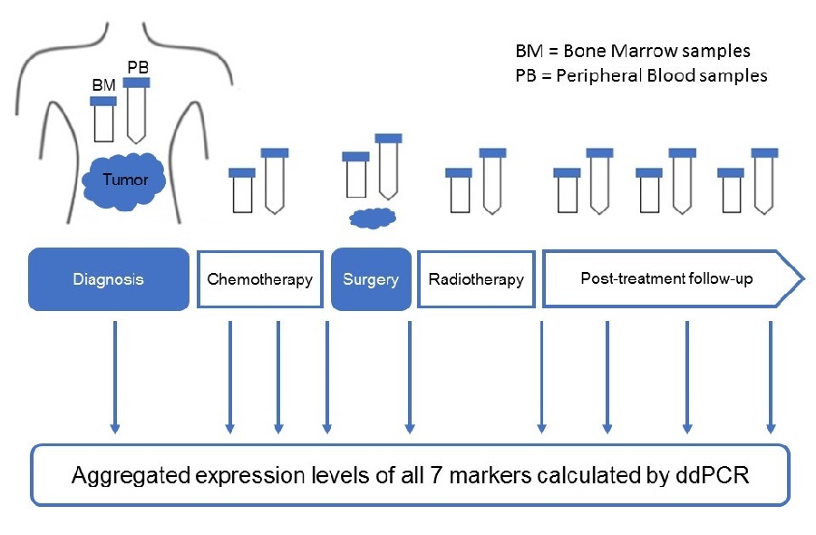
First of all, 208 bone marrow samples and 67 peripheral blood samples were taken from high- risk neuroblastoma patients, and 103 bone marrow samples and 107 peripheral blood samples were obtained from healthy individuals (control). Seven markers were selected from among the eleven markers that are highly expressed in the cancer stem cells that cause neuroblastoma regrowth/relapse. Using ddPCR, they calculated the levels of these seven markers in the bone marrow and peripheral blood samples.
The research group discovered that they could more accurately diagnose high-risk neuroblastoma patients when calculating the aggregated expression levels of all 7 markers using ddPCR, as opposed to looking at the expression level of each marker individually. The changes in the expression levels of these seven markers reflected the amount of tumors, the stage of illness (remission, stable, or progression) and the sample collection period (diagnosis, treatment, post-treatment, or relapse).
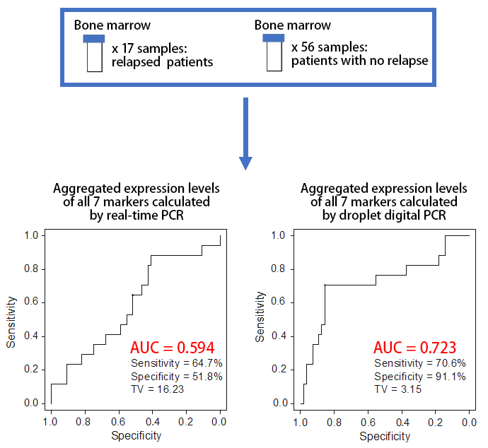
The expression levels of the 7 markers were calculated for 73 bone marrow samples obtained from post-treatment high-risk neuroblastoma patients. The expression levels of the markers in the bone marrow samples of the 17 patients who had suffered tumor relapse/regrowth was significantly higher compared to the levels in the 56 patients who hadn’t. By comparing the two PCR methods using the same 73 samples, it was predicted that the expression levels of the seven markers calculated by ddPCR would be able to more accurately predict tumor relapse/regrowth than qPCR.
Based on these results, it is expected that the ddPCR monitoring method developed by this study will improve the monitoring of MRD in high-risk neuroblastoma patients, enabling the possibility of tumor relapse or regrowth to be more accurately predicted.
Further Developments
The results of this research suggest that it is possible to accurately monitor MRD in post-treatment high-risk neuroblastoma patients. Next, this methodology needs to be evaluated through prospective clinical studies on a larger number of patients across multiple hospitals. It is expected that this monitoring method can be used as a basis for predicting patients’ prognoses and developing new treatments for high-risk neuroblastoma patients.
Glossary
- *1. Droplet digital PCR (ddPCR)
- PCR is a technique used to measure the amount of target DNA or RNA in a sample. In ddPCR, the sample is divided into a large number of individual drops prior to the PCR reaction. Compared to real-time PCR, ddPCR is: 1. More sensitive (it can be used to detect extremely low levels of the target), and 2. Reproducible (unlike qPCR it doesn’t rely on a calibration curve). It is known as the third generation PCR technique.
- *2. Real-time PCR (qPCR)
- This PCR enables the detection of amplified DNA or RNA to be monitored and analyzed in real time. It is known as the second generation PCR technique.
- *3. AUC (Area Under the Curve)
- A ROC (Receiver Operating Characteristic) curve is used for accurate data evaluation and to compare new data. The AUC (Area Under the Curve) is the area underneath a ROC curve on a graph. The AUC has a value between 0 and 1; the closer the value is to 1, the higher the prognosis and diagnosis ability.
Journal Information
- Title
- "Level of seven neuroblastoma-associated mRNAs detected by droplet digital PCR is associated with tumor relapse/regrowth of high-risk neuroblastoma patients"
- DOI
- 10.1016/j.jmoldx.2019.10.012
- Authors
- Khin Kyae Mon Thwin*1, Toshiaki Ishida*2, Suguru Uemura*1, Nobuyuki Yamamoto*1, Kyaw San Lin*1, Akihiro Tamura*2, Aiko Kozaki*2, Atsuro Saito*2, Kenji Kishimoto*2, Takeshi Mori*2, Daiichiro Hasegawa*2, Yoshiyuki Kosaka*2, Nanako Nino*1, Satoru Takafuji*1, Kazumoto Iijima*1, and Noriyuki Nishimura*3
*1 : Department of Pediatrics, Kobe University Graduate School of Medicine
*2 : Department of Hematology and Oncology, Kobe Children’s Hospital
*3 : Department of Public Health, Kobe University Graduate School of Health Science - Journal
- Journal of Molecular Diagnostics