Researchers at Kobe University’s Graduate School of Medicine have developed the world’s first congenital pituitary hypoplasia (CPH) (*1,*2) model using patient-derived iPS cells. The research group consisting of Associate Professor TAKAHASHI Yutaka, medical researcher MATSUMOTO Ryusaku and Professor AOI Takashi et al. succeeded in using the model to illuminate the mechanisms underlying CPH. The team has already been attempting to apply this model to other pituitary diseases and drug discovery.
The results of this study were published in the American Scientific Journal ‘J Clinical Investigation’ on December 17, 2019.
Main points
- Successfully created the world’s first model of pituitary disease using human iPS cells.
- iPS cells were generated from a patient with CPH. These iPS cells enabled the recapitulation of the disease in vitro; an impaired differentiation into the pituitary.
- The causal gene mutation was identified in the patient by exome sequencing analysis (*3). Using this disease model, it was revealed that a deficiency in the FGF10 produced in the neighboring hypothalamus (*4) caused CPH.
- Expected application of this model to other pituitary diseases, leading to the clarification of the underlying mechanisms and drug discovery.
Research Background
Hypopituitarism caused by CPH is not uncommon and it is sometimes life-threating. Patients with this disease require lifelong hormone replacement therapy. The causes and underlying mechanisms are not well understood.
Prior research on pituitary diseases has been mainly conducted using animal models, such as knock out mouse (*5). However, sometimes there are differences in the phenotypes between animal and human. This means that human models are necessary in order to fully understand the mechanisms of such diseases.
In recent years, iPS (induced pluripotent stem) cells have been utilized in the development of disease models, regenerative medicine, and drug discovery. In addition, a method using iPS cells to induce differentiation of both the pituitary and hypothalamus in vitro has been developed; however it had yet to be applied to pituitary disease modeling.
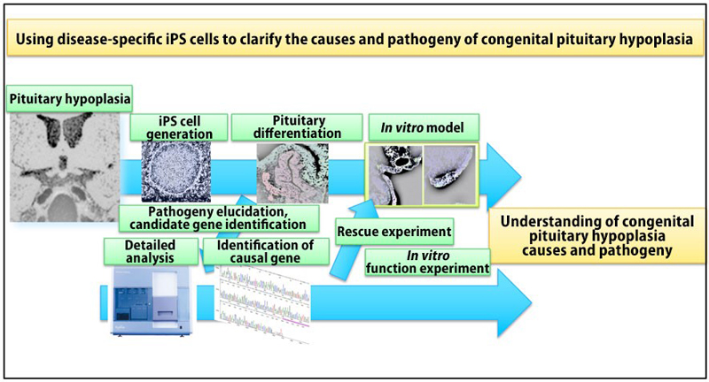
With this in mind, researchers at Kobe University’s Graduate School of Medicine have applied this strategy to develop a human model of CPH in vitro using iPS cells to understand the pathophysiology and causes of the disease (Figure 1).
Research Methodology
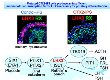
iPS cells were generated from blood samples taken from a patient with CPH. This patient exhibited congenital pituitary hypoplasia and was undergoing pituitary hormone replacement therapy. These patient-derived iPS cells were utilized to elucidate the underlying mechanisms in vitro. Interestingly, control iPS cells differentiated into hormone-producing cells, however, the CPH patient-derived iPS cells were not able to differentiate into these cells. Further analysis of the differentiation process revealed that the transcription factor LHX3, which is essential for pituitary differentiation, was not expressed in the pituitary progenitor from patient-derived iPS cells (Figure 2). Exome sequencing analysis revealed a mutation in the OTX2 gene and that its function was impaired. Correction of the OTX2 mutation in patient-derived iPS cells restored the pituitary differentiation ability, demonstrating that the OTX2 mutation was responsible.
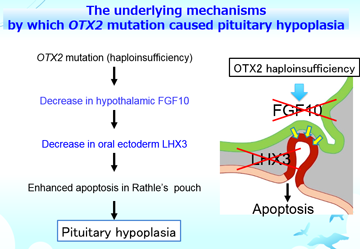
Interactions between the pituitary and the adjacent hypothalamus are essential for pituitary differentiation and regulation. An advantage of this model is that it can simultaneously develop the pituitary and hypothalamus in vitro from iPS cells. This allowed the researchers to investigate the significance of interaction between these tissues. They clarified that the hypothalamus was responsible for the disease by performing chimera formation experiments using both patient-derived iPS cells and healthy iPS cells. Subsequent analysis exhibited that FGF10 (fibroblast growth factor 10) from the hypothalamus plays a pivotal role in the expression of the transcription factor LHX3 in the pituitary. Furthermore, expression levels of LHX3 were restored by adding FGF10 in vitro. These results demonstrated that FGF10 deficiency in the hypothalamus associated with OTX2 mutation was responsible.
Collectively, the OTX2 mutation caused a decrease in hypothalamic FGF10, resulting in a lack of LHX3 expression in the oral ectoderm, which is the precursor of the pituitary. Consequently, loss of LHX3 caused apoptosis of the precursor cells, therefore causing the impaired development of the pituitary. These underlying mechanisms were illuminated for the first time by this study (Figure 3).
This pituitary disease model utilizing human iPS cells has elucidated the detailed underlying mechanisms, which animal models were unable to reveal.
Further Research
This research revealed the pathophysiology of CPH through disease-specific iPS cells (*6). Furthermore, the model was also useful for understanding the pituitary differentiation process in humans.
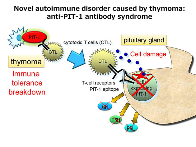
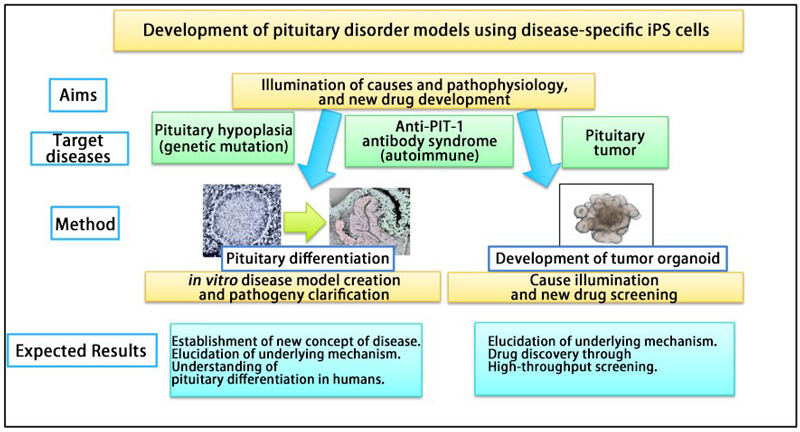
This research team is also investigating the causes, pathogenesis and treatment of other pituitary diseases (such as autoimmune disorders and pituitary tumors) using the same strategy. In particular, they are continuing to develop pituitary disease models to investigate the pathophysiology and precise mechanisms underlying the ‘Anti-PIT-1 antibody syndrome’ (*7), which is a novel type of autoimmune pituitary disease that the team has established as a new clinical entity. Using this model, they have already demonstrated the antigen presentation of PIT-1 protein epitope on the anterior pituitary cells derived from patient iPS cells.
In addition, there are many other incurable pituitary diseases, for which the causes remain unknown. It is hoped that these iPS cell-based research methods for the diseases will lead to the clarification of the underlying mechanisms and drug discovery.
Glossary
- *1. Pituitary
- Endocrine gland located at the base of the brain. It consists of the posterior and anterior pituitary and is responsible for secreting various vital hormones.
- *2. Congenital Pituitary Hypoplasia
- A disorder in which the pituitary isn’t formed correctly due to genetic mutation. Lifelong hormone treatment is necessary for many patients.
- *3. Exome sequencing analysis
- A technique that allows all the protein-coding regions of genes in the human genome to be analyzed.
- *4. Hypothalamus
- Located near the pituitary, the hypothalamus plays an important role in many functions, including regulating body temperature and releasing hormones. In the early stages of development, the hypothalamus controls various factors relating to pituitary differentiation. Hypothalamic hormones also regulate hormones produced by the pituitary gland.
- *5. Knock-out mouse
- allows the function of a particular gene to be researched. The gene of interest is removed from the mouse using genetic engineering.
- *6. Disease-specific iPS cells
- These are iPS cells generated from a patient that has a particular disease. They can be used to understand the causes behind various diseases and in the development of new treatments.
- *7. Anti-PIT-1 antibody syndrome
- A disease discovered by the members of current research team that leads to acquired hypopituitarism. This syndrome is caused by an autoimmunity to the transcription factor PIT-1 and is characterized by deficiencies in GH, TSH and PRL. The transcription factor PIT-1 is essential for the differentiation and maintenance for these hormone-producing cells. In anti-PIT-1 antibody syndrome, cytotoxic T cells destroy cells expressing PIT-1, thus causing the specific damage in pituitary gland. The underlying mechanism underlying the disease, involving thymomas, has yet to be fully illuminated.
- *8. Epitope
- part of an antigen that is recognized by the immune system.
Acknowledgements
Professor TAKAHASHI would like to express his deep gratitude towards his co-researchers; Dr. SUGA Hidetaka (Nagoya University), Professor HASEGAWA Tomonobu (Keio University), Dr. NARUMI Satoshi (National Center for Child Health and Development), Professor MUGURUMA Keiko (Kansai Medical University) and Professor OGAWA Wataru (Kobe University).
This research was supported by grants from AMED ( ‘The Acceleration Program for Intractable Disease Research Utilizing Disease-specific iPS Cells’ ), MEXT, JSPS, the Uehara Memorial Foundation, and the Naito Foundation.
Journal Information
- Title
- “Congenital pituitary hypoplasia model demonstrates hypothalamic OTX2 regulation of pituitary progenitor cells”
- DOI
- 10.1172/JCI127378
- Authors
- Ryusaku Matsumoto, Hidetaka Suga, Takashi Aoi, Hironori Bando, Hidenori Fukuoka, Genzo Iguchi, Satoshi Narumi, Tomonobu Hasegawa, Keiko Muguruma, Wataru Ogawa, Yutaka Takahashi
- Journal
- J Clinical Investigation