An international research group including Professor Kaoru Sugasawa of the Biosignal Research Center, Kobe University, Dr. Fumio Hanaoka, Director-General of the National Institute of Genetics, and Dr. Wei Yang of the National Institutes of Health, USA, has successfully elucidated the elaborate molecular mechanism that ensures the proper repair of DNA damaged by ultra-violet light or chemical carcinogens. This work is expected to deepen our understanding of DNA repair mechanisms that prevent cancer and other diseases. The results of this research were published in Nature at 16:00 UTC on Wednesday, April 19th.
Main Points
- XPC protein acts as a sensor to detect various abnormalities in the DNA structure.
- The faithful repair of DNA is ensured by using the basal transcription factor TFIIH and XPA protein to verify the presence of any lesion that needs to be repaired.
- Using the XPB and XPD proteins present in TFIIH as “motors” to pull the DNA into the protein complex from both directions, the presence of damage is finally verified when the movement of a DNA strand is blocked by the lesion.
- The present research provides the first explanation of the detailed function and mechanism of xeroderma pigmentosum causative gene products, such as XPA, XPB and XPD, in DNA repair processes.
Research Background
Our genomic DNA is continuously damaged by endogenous factors such as reactive oxygen species, and also by environmental factors such as ultra-violet light, radiation, and chemicals. Failure to repair damaged DNA may induce mutations and cell death, eventually leading to the onset of cancer and other diseases. To prevent this, our cells are equipped with various defense systems aimed at finding and repairing damaged DNA.
Nucleotide excision repair (NER) is an important mechanism for the repair of diverse DNA lesions caused by ultra-violet light and chemical carcinogens. Many patients diagnosed with xeroderma pigmentosum (XP) have mutations in one of the genes encoding proteins involved in the NER mechanism. It is known that their high susceptibility to ultra-violet light induced skin cancer in particular is due to the deficiency in NER function. This also tells us that NER serves as a defense against cancer.
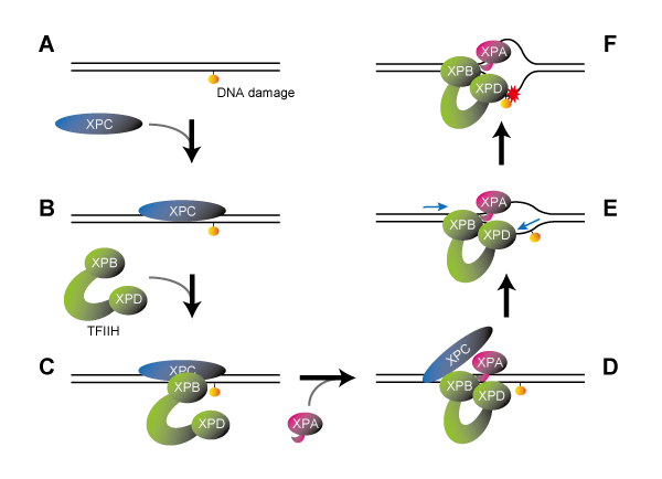
The first step of NER involves the XPC protein, one of the several XP causative gene products, which operates as a “sensor” to locate abnormalities in the DNA structure. XPC has a potential to deal with a wide variety of DNA lesions, while it also binds to DNA sites without any damage, such as base mismatches. Since unnecessary repair reactions at such improper sites may increase a risk of adverse mutations, it is essential to verify if there is any damage that needs to be repaired. Although it has previously been shown that the basal transcription factor TFIIH and XPA protein play a role in such a damage verification step, the detailed mechanism has remained unclear up to now.
Research Methodology
Following the known reaction steps in the NER process, the current researchers prepared a series of complexes containing damaged DNA sequentially bound by XPC, TFIIH and XPA, which were then subjected to cryogenic electron microscopy to analyze their molecular structures in detail. TFIIH is a large protein complex composed of seven subunits, including XPB and XPD proteins, which plays crucial roles in both NER and transcription (gene expression). It usually adopts a U-shaped “horseshoe” structure, with the XPB and XPD proteins positioned at the tips of its two open arms. This horseshoe structure was retained in the DNA-XPC-TFIIH complex, with XPB bound to XPC at the damage site, while XPD does not interact with either DNA or XPC (Figure 1C). When XPA was subsequently involved, however, TFIIH underwent a major conformational change. While the two open arms of the horseshoe came together to form a ring-shaped structure, XPB moved away from the damaged site such that XPD was allowed to bind DNA in between (Figure 1D).
Credit: SUGASAWA Kaoru, License:CC BY
How does this molecular arrangement then verify the presence of DNA damage? It is known that XPB functions as a "DNA translocase", which binds to the DNA duplex and move along one of the DNA strands. Based on the newly elucidated structure for the complex, it was assumed that XPB should move along the DNA away from the damage site. However, since it is constrained by other proteins, XPB cannot itself move actually: instead, the DNA duplex moves in the opposite direction. In other words, XPB pushes DNA into the complex (Figure 1E). Because the structure revealed that part of XPA is inserted between the two DNA strands behind XPB, this is expected to unwind the DNA duplex into single strands like a fastener being unzipped. Moreover, it was also known that XPD binds to single-stranded DNA and move along it in a defined direction (5’→3’). Of the two DNA strands unraveled by XPB and XPA, XPD binds to the damaged one. However, as is the case with XPB, the XPD cannot itself move so the damaged strand is likewise pulled towards the complex (Figure 1E). When bound to a single-stranded DNA, a narrow pinhole is formed in XPD, through which the DNA strand passes. Therefore, if there is a bulky lesion on the DNA strand, that part will be trapped at the entrance to XPD such that the strand will stop its moving (like a knotted thread being unable to pass through the eye of a needle: Figure 1F). The present research thus successfully elucidated the mechanism whereby the presence of a DNA lesion is verified: the decision to proceed with the repair reaction is made by the blockage of DNA strand movement through XPD. In fact, previous researches have indicated that non-bulky DNA lesions (base detachment, etc.) that could be predicted to pass through the XPD hole are not a target for the NER repair process.
Further Research
The current research has clarified which parts of proteins such as XPA, XPB, and XPD are involved in the NER process to repair damaged DNA together with their mechanism of action. As our understanding deepens about the negative impact on repair response arising from structural changes induced in pathogenic variants of these proteins in XP patients, it may also be possible to support the future development of drugs and other treatments. The fact that Japan in particular has an overwhelmingly high ratio of XP patients with variant XPA, places huge significance on improving our understanding of the relationship between protein structure and function.
Glossary
Xeroderma pigmentosum (XP)
A recessive hereditary disorder in humans characterized by hypersensitivity to sunlight and a high incidence of skin cancer. XP patients have biallelic mutations in one of eight types of causative genes. XPA –XPG proteins, expressed from seven of these genes (XPA – XPG), are directly involved in the nucleotide excision repair (NER). Designated as an intractable disease, at least half the XP patients in Japan are attributed to variant XPA genes.
Cryogenic electron microscopy
A microscopy method that analyzes biological samples at low temperatures (usually -196ºC, liquid nitrogen), causing much lower electron beam damage to the sample than by conventional transmission electron microscopy. This method allows the observation of high-resolution structure by maintaining the sample condition closer to that within the living organism. Awarded the Nobel Prize in Chemistry in 2017.
Acknowledgements
This study was supported by Grants-in-Aid for Scientific Research (S) (JP16H06307) and (B) (JP21H03598), from the Japan Society for the Promotion of Science.
Journal Information
Title
“Lesion recognition by XPC, TFIIH and XPA in DNA excision repair”
DOI
10.1038/s41586-023-05959-z
Authors
: Jinseok Kim, Chia-Lung Li, Xuemin Chen, Yanxiang Cui, Filip M. Golebiowski, Huaibin Wang,Fumio Hanaoka, Kaoru Sugasawa, and Wei Yang
Journal
Nature